Our Work
Pancreatic Cysts
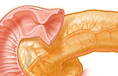
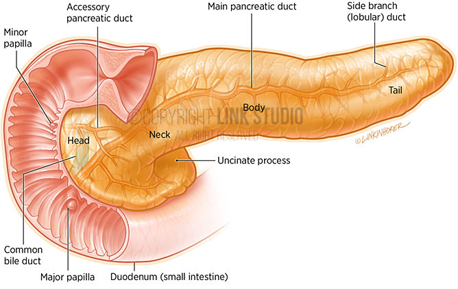
Pancreatic Anatomy
Pancreatic cystic lesions may develop within or be associated with various anatomical structures of the pancreas, such as the pancreatic ducts.
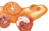
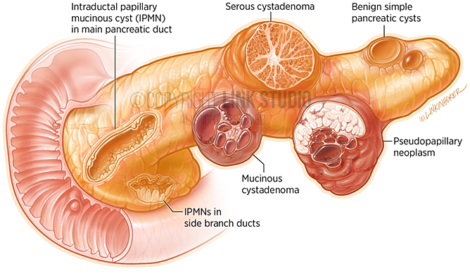
Cystic Lesions of the Pancreas
Cystic lesions of the pancreas are benign or malignant masses that come in a variety of forms. These masses need to be identified so to understand the malignant potential of the lesion. This often requires long-term surveillance and the review of endoscopic ultrasound images, lab work, and other radiograph images.
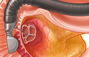
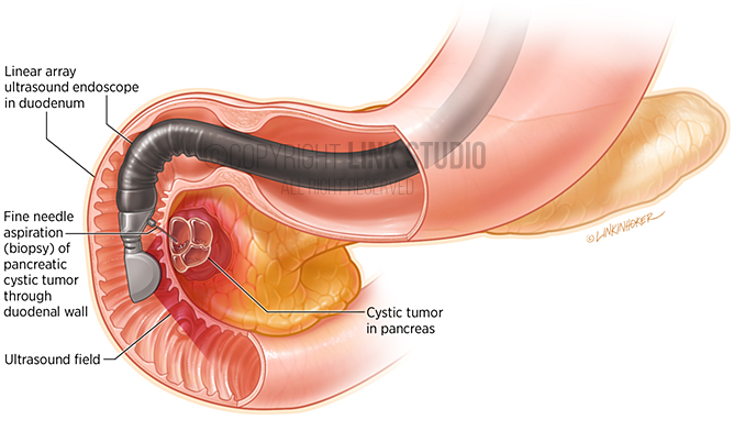
Endoscopic Ultrasound with Fine Needle Aspiration (EUS-FNA) of a Pancreatic Cyst
Endoscopic ultrasound (EUS) is used to view and better diagnose cystic lesions in the pancreas. A specialized endoscope is placed in the upper gastrointestinal tract, next to the pancreas, allowing proper examination. The EUS is also used to guide fine needle aspiration (removal) of fluid and solid material within the lesion.
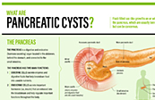

Infographic: What are Pancreatic Cysts?
The infographic describes the importance of the pancreas, where it is located in the body, what pancreatic cysts and pseudocysts are, where they come from, and how pancreatic cysts are diagnosed.