Our Work
The Liver & Hepatitis
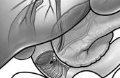
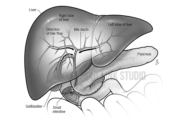
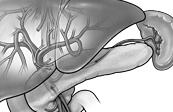
Biliary Drainage of the Liver
Bile is formed and drains into the biliary ducts which eventually forms the right and left hepatic ducts, which join to form the common hepatic duct, after which it combines with the cystic duct to become the common bile duct (CBD). The CBD combines with the pancreatic duct at the ampulla of Vater and drains through the greater papilla into the small intestine.
This series of illustrations is part of the book Hepatitis C, A Complete Guide for Patients and Families.
This illustration is available as Stock | Contact us now for licensing options
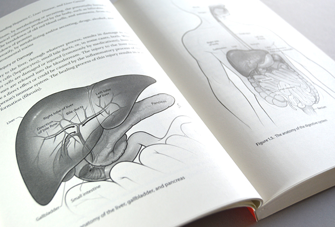
Book Spread – Biliary Drainage of the Liver
Bile is formed and drains into the biliary ducts which eventually forms the right and left hepatic ducts, which join to form the common hepatic duct, after which it combines with the cystic duct to become the common bile duct (CBD). The CBD combines with the pancreatic duct at the ampulla of Vater and drains through the greater papilla into the small intestine.
This series of illustrations is part of the book Hepatitis C, A Complete Guide for Patients and Families.
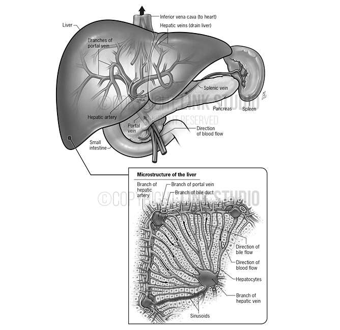
Blood Supply & Drainage of the Liver
This image illustrates the portal vein, which drains all the blood from the intestines through the liver; the hepatic arteries, which supply blood to the liver; and the hepatic veins, which drain the liver. The inset shows the histological (microscopic) structure of liver tissue.
This series of illustrations is part of the book Hepatitis C, A Complete Guide for Patients and Families.
This illustration is available as Stock | Contact us now for licensing options
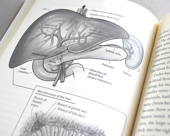
Book Spread – Blood Supply & Drainage of the Liver
This image illustrates the portal vein, which drains all the blood from the intestines through the liver; the hepatic arteries, which supply blood to the liver; and the hepatic veins, which drain the liver. The inset shows the histological (microscopic) structure of liver tissue.
This series of illustrations is part of the book Hepatitis C, A Complete Guide for Patients and Families.
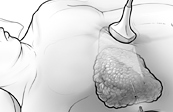
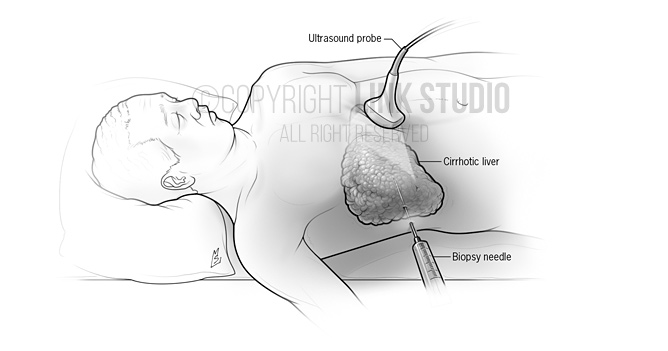
Ultrasound Guided Liver Biopsy of a Cirrhotic Liver
This series of illustrations is part of the book Hepatitis C, A Complete Guide for Patients and Families.
This illustration is available as Stock | Contact us now for licensing options