Our Work
Heart Anatomy
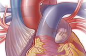
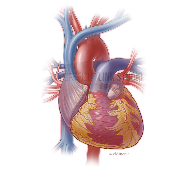
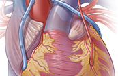
Anterior View of the External Heart
The anterior view focuses on the external structures viewable from the front, such as the four chambers, great vessels, right and left coronary arteries, left anterior interventricular branch, and left circumflex branch.
This illustration is available as Stock | Contact us now for licensing options
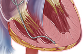
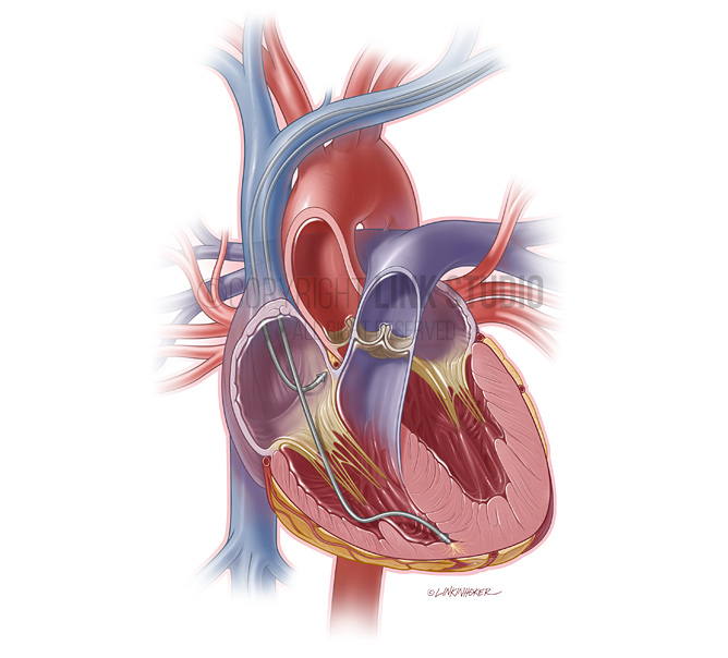
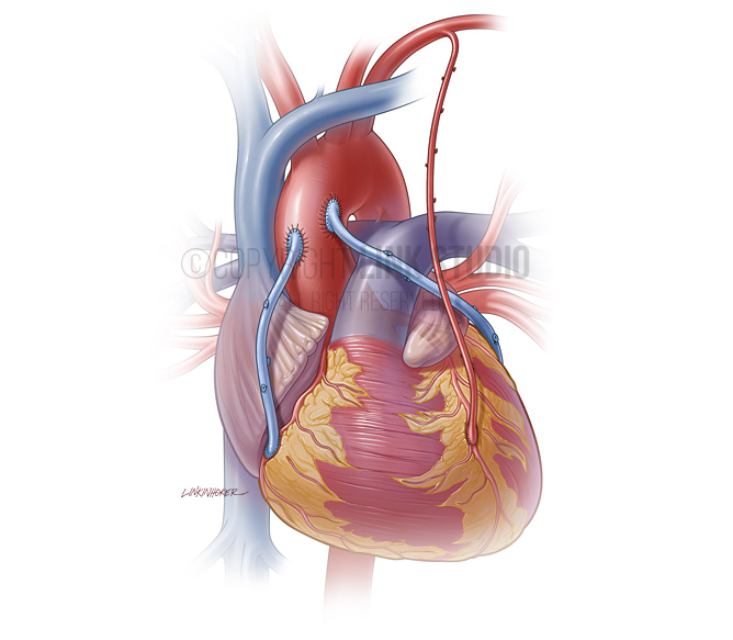
Anterior View of the Internal Heart with Pacemaker Leads
The pacemaker is implanted into the subcutaneous space in the upper chest, while the leads are introduced into the subclavian veins, where one is placed into the right atrium and the other is placed into the apex of the right ventricle. The pacemaker is used to help stabilize abnormal heart rhythms.
This illustration is available as Stock | Contact us now for licensing options
Triple Coronary Artery Bypass Grafts
In this version of bypass surgery, two saphenous vein grafts are used to bypass blockages in the right coronary artery and the circumflex branch of the left coronary artery, while the left internal thoracic artery is used to bypass a blockage in the left anterior descending artery.
This illustration is available as Stock | Contact us now for licensing options