Our Work
Brain Development & Zika Virus
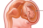
Zika Virus Infection of Apical Radial Glial Cells (aRGC) in the Developing Brain
Zika virus gains entry into neuroprogenitor cells (aRGC) of the developing cerebral cortex by binding to the Axl receptor present on the surface of the aRGCs. Within the aRGCs, the virus divides and vastly increases it population and infectious potential. This illustration accompanies an article introducing the general public, students, and policy makers to the field of brain development, the Zika virus, and infectious disease.
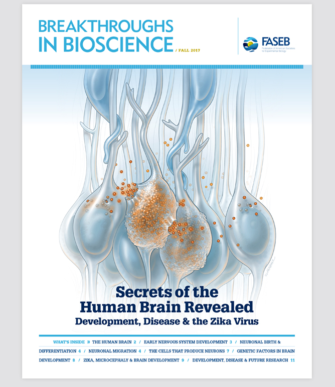
Breakthroughs in Bioscience Cover: Zika Virus Infection of Apical Radial Glial Cells (aRGC) in the Developing Brain
Zika virus gains entry into neuroprogenitor cells (aRGC) of the developing cerebral cortex by binding to the Axl receptor present on the surface of the aRGCs. Within the aRGCs, the virus divides and vastly increases it population and infectious potential. This illustration accompanies an article introducing the general public, students, and policy makers to the field of brain development, the Zika virus, and infectious disease.
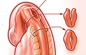
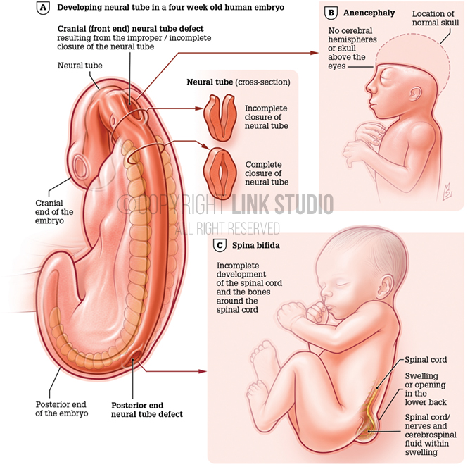
Abnormalities During Neural Tube Development
The neural tube forms from the folding of the neural plate early in neural development. By the end of the fourth week of gestation, the ends of the neural tube close. However, the failure of the neural tube to close during development leads to lethal deformities or lifelong disabilities, such as anencephaly and spina bifida.
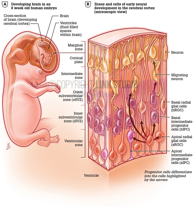
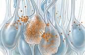
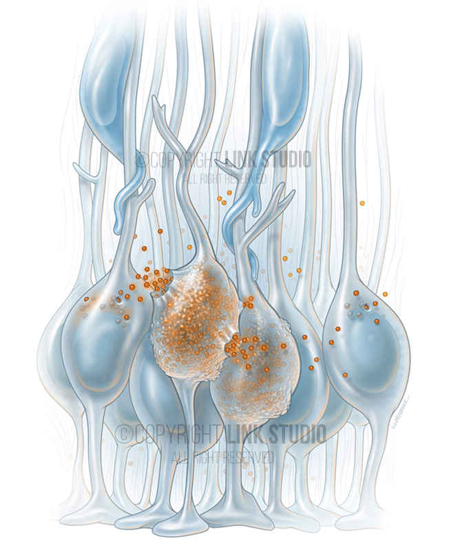
Neural Progenitor Cells in the Developing Cerebral Cortex
Neural progenitor cells proliferate and differentiate, giving rise to multiple types of cortical cells of the developing brain.