Our Work
3D Bioprinting


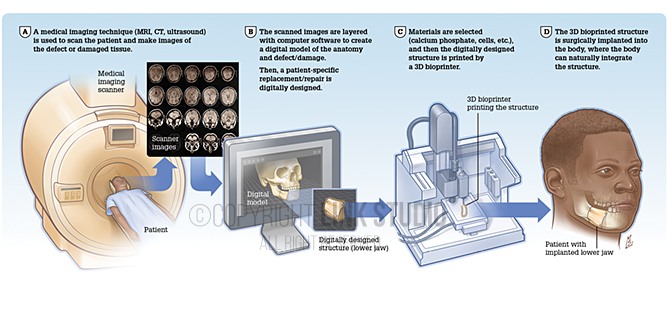
3D Bioprinting Process for a Mandible (Lower Jaw) Defect
A medical imaging technique (MRI, CT, ultrasound) is used to scan the patient and make images of the defect or damaged tissue. Next, The scanned images are layered with computer software to create a digital model of the anatomy and defect/damage. Then a patient-specific replacement/repair is digitally designed. After which materials are selected (calcium phosphate, cells, etc.) and then the digitally designed structure is printed by a 3D bioprinter. Finally, the 3D bioprinted structure is surgically implanted into the body, where the body can naturally integrate the structure.


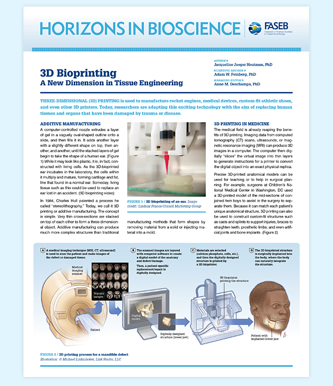
Horizons In Bioscience Publication – 3D Bioprinting, A New Dimension in Tissue Engineering
Redesign for the FASEB publication that describes scientific discoveries on the brink of clinical application for a wide ranging audience, including policy makers, students, and the general public.
Client
- Federation of American Societies for Experimental Biology (FASEB)
Category
- Medical Illustration
Industry
- Publishing
- Academic
- Pharma / Biotechnology
Audience
- General Public
- Patient Education
Specialty
- Biotechnology